Attēls:Bronchiolar epithelium 3 - SEM.jpg

Šī priekšskata izmērs: 585 × 599 pikseļi. Citi izmēri: 234 × 240 pikseļi | 469 × 480 pikseļi | 750 × 768 pikseļi | 1 024 × 1 049 pikseļi.
Sākotnējais fails (1 024 × 1 049 pikseļi, faila izmērs: 375 KB, MIME tips: image/jpeg)
| Šis fails ir no Vikikrātuves. Tā apraksts no attēla lapas Vikikrātuvē ir parādīts zemāk. Vikikrātuve ir brīvi licencēta failu krātuve. Tu vari tai palīdzēt. |
Kopsavilkums
| Apraksts |
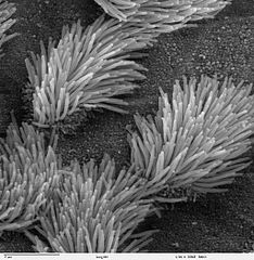
Scanning electron microscope image of lung trachea epithelium. There are both ciliated and non-ciliated cells in this epithelium. Note the difference in size between the cilia and the microvilli (on the non-ciliated cell surface). Zeiss DSM 962 SEM |
| Avots | |
| Autors | Charles Daghlian |
| Atļauja: (Šī faila izmantošana citur) |
PD |
Licence
| This work has been released into the public domain by its author, Charles Daghlian. This applies worldwide. In some countries this may not be legally possible; if so: Charles Daghlian grants anyone the right to use this work for any purpose, without any conditions, unless such conditions are required by law.
|
Faila hronoloģija
Uzklikšķini uz datums/laiks kolonnā esošās saites, lai apskatītos, kā šis fails izskatījās tad.
| Datums/Laiks | Attēls | Izmēri | Dalībnieks | Komentārs | |
|---|---|---|---|---|---|
| tagadējais | 2006. gada 7. oktobris, plkst. 17.16 | | 1 024 × 1 049 (375 KB) | Patho | {{Information |Description=Scanning electron microscope image of lung trachea epithelium. There are both ciliated and on-ciliated cells in this epithelium. Note the difference in size between the cilia and the microvilli(on non-ciliated cell surface) Zei |
Faila lietojums
Šo failu izmanto šajā 1 lapā:
Globālais faila lietojums
Šīs Vikipēdijas izmanto šo failu:
- Izmantojums ar.wikipedia.org
- Izmantojums ast.wikipedia.org
- Izmantojums bs.wikipedia.org
- Izmantojums ca.wikipedia.org
- Izmantojums cs.wikipedia.org
- Izmantojums da.wikipedia.org
- Izmantojums de.wikipedia.org
- Izmantojums de.wikibooks.org
- Izmantojums en.wikipedia.org
- Izmantojums es.wikipedia.org
- Izmantojums eu.wikipedia.org
- Izmantojums fa.wikipedia.org
- Izmantojums fr.wikipedia.org
- Izmantojums gl.wikipedia.org
- Izmantojums he.wikipedia.org
- Izmantojums he.wiktionary.org
- Izmantojums hi.wikipedia.org
- Izmantojums id.wikipedia.org
- Izmantojums jv.wikipedia.org
- Izmantojums kk.wikipedia.org
- Izmantojums lt.wikipedia.org
- Izmantojums ms.wikipedia.org
- Izmantojums nl.wikipedia.org
- Izmantojums nn.wikipedia.org
- Izmantojums no.wikipedia.org
- Izmantojums pl.wikipedia.org
- Izmantojums pl.wiktionary.org
- Izmantojums pt.wikipedia.org
- Izmantojums ru.wikipedia.org
- Izmantojums ru.wiktionary.org
- Izmantojums sh.wikipedia.org
- Izmantojums simple.wikipedia.org
Skatīt šī faila pilno globālo izmantojumu.