Attēls:Anaplastic astrocytoma.jpg
Šī priekšskata izmērs: 800 × 552 pikseļi. Citi izmēri: 320 × 221 pikseļi | 640 × 442 pikseļi | 1 024 × 707 pikseļi | 1 200 × 828 pikseļi.
Sākotnējais fails (1 200 × 828 pikseļi, faila izmērs: 200 KB, MIME tips: image/jpeg)
| Šis fails ir no Vikikrātuves. Tā apraksts no attēla lapas Vikikrātuvē ir parādīts zemāk. Vikikrātuve ir brīvi licencēta failu krātuve. Tu vari tai palīdzēt. |
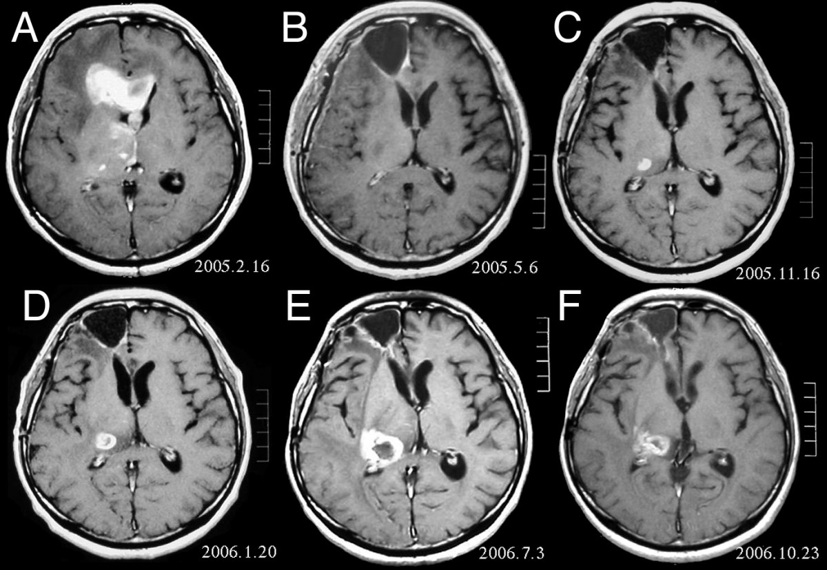
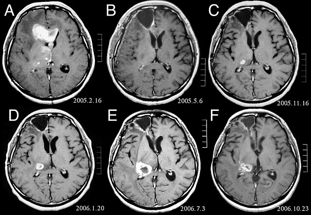
| Apraksts | MRI of brain. (A) Initial MRI on February 16, 2005, shows a tumor in the right and left frontal lobe as well as the right thalamus. (B) MRI after surgery, radiation and chemotherapy. The tumor has completely disappeared except for slight enhancement adjacent to the surgical margin. (C) Recurrence of the thalamic tumor despite maintenance chemotherapy on November 16, 2005. (D) Increase in size of the thalamic tumor two months after stereotactic radiotherapy. (E) After 6 cycles of TMZ therapy, the thalamic lesion enlarged, and the patient developed dysarthria and hemiparesis. (F) After 2 courses of treatment with interferon-beta and TMZ, the tumor shows a partial response. |
| Datums | |
| Avots | Fujimaki T, Ishii H, Matsuno A, Arai H, Nakagomi T.Effectiveness of interferon-beta and temozolomide combination therapy against temozolomide-refractory recurrent anaplastic astrocytoma.World J Surg Oncol. 2007 Aug 4;5:89. PMID 17683572 doi:10.1186/1477-7819-5-89 |
| Autors | Fujimaki T, Ishii H, Matsuno A, Arai H, Nakagomi T. |
| Atļauja: (Šī faila izmantošana citur) |
BioMedCentral License |
Šis fails tiek izplatīts saskaņā ar licences Creative Commons Atsauce 2.0 Vispārējiem noteikumiem.
- Jūs varat brīvi:
- koplietot – kopēt, izplatīt un pārraidīt darbu
- remiksēt – pielāgot darbu
- Saskaņā ar šādiem nosacījumiem:
- atsaucoties – Tev ir jānorāda autors, saite uz licenci un to, vai veiktas kādas izmaiņas. To var darīt jebkādā saprātīgā veidā, bet ne tādā, kas norādītu, ka licencētājs atbalsta tevi vai veidu, kā tu izmanto šo darbu.
Faila hronoloģija
Uzklikšķini uz datums/laiks kolonnā esošās saites, lai apskatītos, kā šis fails izskatījās tad.
| Datums/Laiks | Attēls | Izmēri | Dalībnieks | Komentārs | |
|---|---|---|---|---|---|
| tagadējais | 2008. gada 25. februāris, plkst. 19.47 | | 1 200 × 828 (200 KB) | Filip em | {{Information |Description=MRI of brain. (A) Initial MRI on February 16, 2005, shows a tumor in the right and left frontal lobe as well as the right thalamus. (B) MRI after surgery, radiation and chemotherapy. The tumor has completely disappeared except f |
Faila lietojums
Šo failu izmanto šajā 1 lapā:
Globālais faila lietojums
Šīs Vikipēdijas izmanto šo failu:
- Izmantojums ar.wikipedia.org
- Izmantojums bg.wikipedia.org
- Izmantojums cs.wikipedia.org
- Izmantojums da.wikipedia.org
- Izmantojums de.wikipedia.org
- Izmantojums el.wikipedia.org
- Izmantojums en.wikipedia.org
- Izmantojums es.wikipedia.org
- Izmantojums et.wikipedia.org
- Izmantojums hi.wikipedia.org
- Izmantojums hr.wikipedia.org
- Izmantojums hu.wikipedia.org
- Izmantojums it.wikipedia.org
- Izmantojums kk.wikipedia.org
- Izmantojums lb.wikipedia.org
- Izmantojums mk.wikipedia.org
- Izmantojums mt.wikipedia.org
- Izmantojums nl.wikipedia.org
- Izmantojums no.wikipedia.org
- Izmantojums pl.wikipedia.org
- Izmantojums ro.wikipedia.org
- Izmantojums sk.wikipedia.org
- Izmantojums sl.wikipedia.org
- Izmantojums sq.wikipedia.org
- Izmantojums sr.wikipedia.org
- Izmantojums uk.wikipedia.org